
Mallet finger
What it is
It’s that condition in which the last part of the finger (distal phalanx) loosee part of its extension. This is caused by an isolated lesion of the extensor tendon (fig.1) or to a fracture of the bone where the same tendon is inserted (fig. 2). This condition could be caused by an important trauma, a sit happens in some sport activities, or to a minor one (such as during daily home activities). The diagnosis is done on clinical examination and on an X ray (to check bony fragment).
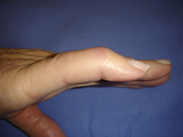
fig. 1
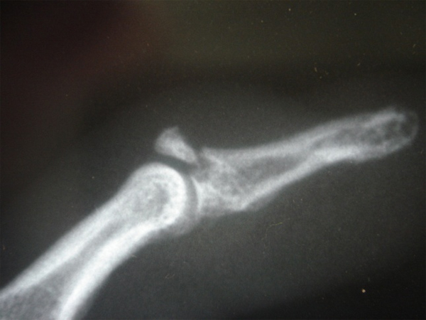
fig. 2

fig. 3
If it is determined that the injury is restricted to the tendon the most effective treatment is to put the finger in a special splint specifically designed. The splinting period lasts for six weeks, after which point you are advised to wear it at night for an additional two weeks. (fig. 3)
When flexion is important or a fracture occurs, surgery becomes necessary to reattach both the bone and the tendon. In this case, fine wires are inserted into the bone to keep it in place as it heals, which will then be removed once the bone has knitted together. Mandatory in both treatment is the postrehabilitative protocol.